Unveiling the Wonders of Leifson Staining Method in Flagella Studies
Flagella, the whip-like appendages protruding from the surface of many microorganisms, play a pivotal role in their movement and interaction with their environment. Understanding the structure and distribution of flagella is crucial in various fields, including microbiology, ecology, and medical research. Among the array of staining techniques available for flagella visualization, the Leifson staining method stands out for its simplicity, effectiveness, and reliability.
The Significance of Flagella Staining
Flagella staining techniques are indispensable tools in microbiology laboratories, enabling researchers to visualize and characterize these essential structures. By staining flagella, microbiologists can distinguish between different types of microorganisms based on their flagellar arrangements, aiding in taxonomic classification. Moreover, flagella staining facilitates the study of bacterial motility, an important trait in pathogenesis, environmental adaptation, and symbiotic relationships.
Introduction to the Leifson Staining Method
The Leifson staining method, developed by Harold Leifson in the mid-20th century, offers a straightforward yet effective approach to stain flagella. This technique is particularly renowned for its ability to accentuate flagellar structures while minimizing background staining, providing clear and precise visualization under a microscope.
Procedure
The Leifson staining method involves a series of simple steps:
1. Preparation of Smear: A bacterial smear is prepared on a glass slide from a fresh culture using aseptic techniques.
2. Fixation: The smear is heat-fixed by passing the slide over a flame or by placing it on a hot plate. Heat fixation helps to adhere the bacterial cells to the slide and denature their proteins, facilitating staining.
3. Staining: The smear is flooded with Leifson’s stain, a combination of basic fuchsin, phenol, and malachite green. The stain penetrates the bacterial cells and selectively binds to the flagella, imparting them a distinct color.
4. Rinsing: Excess stain is rinsed off using distilled water to remove any unbound dye and reduce background staining.
5. Mounting: A coverslip is placed over the stained smear, using a mounting medium such as glycerol to prevent drying and preserve the sample.
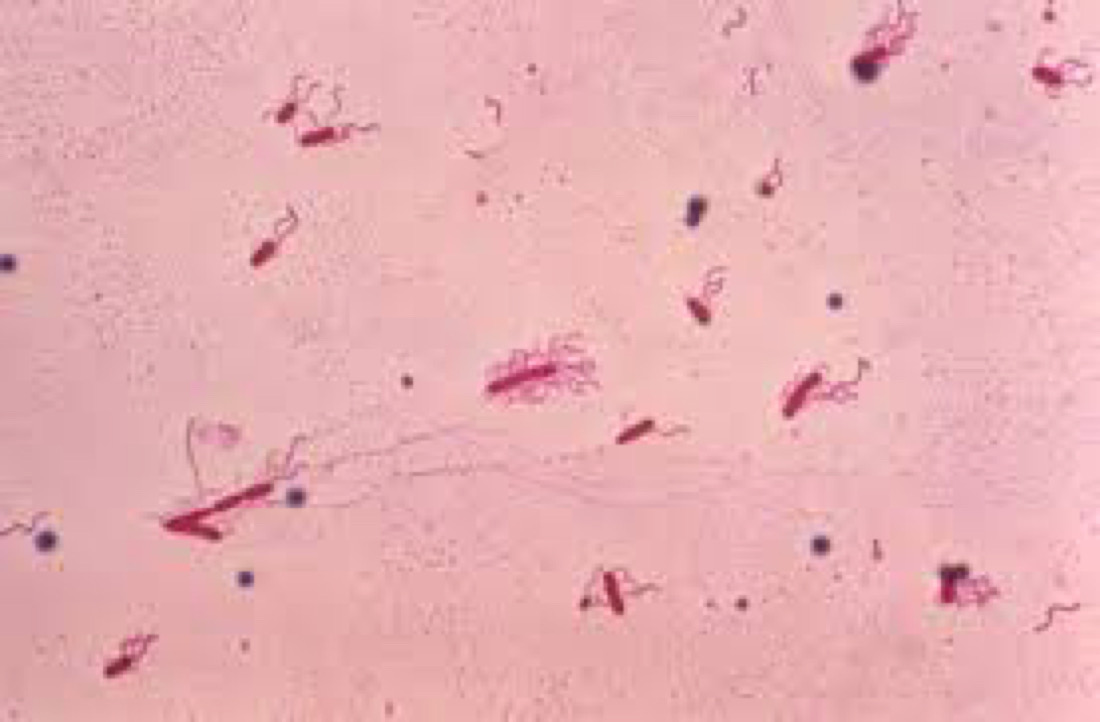
6. Examination: The stained slide is examined under a light microscope using oil immersion at high magnification. Flagella appear as slender, thread-like structures protruding from the bacterial cells, allowing for detailed observation and analysis.
Advantages of Leifson Staining Method
The Leifson staining method offers several advantages over other flagella staining techniques:
1. Simplicity: The procedure is relatively simple and can be performed with basic laboratory equipment, making it accessible to researchers with varying levels of expertise.
2. Clarity: Leifson staining provides clear and distinct visualization of flagella, allowing for accurate interpretation and analysis of their distribution and arrangement.
3. Specificity: The stain selectively binds to flagellar structures, minimizing non-specific background staining and enhancing contrast between the flagella and the bacterial cells.
4. Versatility: The Leifson staining method can be adapted for use with a wide range of bacterial species, making it a versatile tool for flagella studies across diverse microbial taxa.
Applications in Microbiology
The Leifson staining method finds widespread application in various fields of microbiology:
– Taxonomy and Classification: Flagella staining facilitates the identification and classification of bacterial species based on their flagellar arrangements, aiding in microbial taxonomy and phylogenetic studies.
– Motility Assays: By visualizing flagella, researchers can assess the motility of bacterial cells, providing insights into their behavior and ecological roles in diverse environments.
– Pathogenesis Studies: Understanding the role of flagella in bacterial pathogenesis is crucial for developing strategies to combat infectious diseases. The Leifson staining method enables researchers to investigate the role of flagella in virulence and host-pathogen interactions.
Conclusion
The Leifson staining method continues to be a cornerstone technique in flagella studies, offering a simple yet powerful approach for visualizing and analyzing these crucial microbial structures. Its versatility, clarity, and reliability make it an indispensable tool in microbiology laboratories worldwide, facilitating research across various disciplines and contributing to our understanding of microbial diversity, behavior, and pathogenesis. As technology advances, new methods may emerge, but the enduring utility of Leifson staining underscores its enduring importance in the study of flagella and microbial biology.


