Unveiling the Microscopic World: Exploring Negative Staining Techniques
Introduction:
Microscopy has been a cornerstone in scientific research, allowing scientists to explore the intricate details of the microscopic world. One technique that has played a crucial role in this endeavor is negative staining. Negative staining is a versatile method employed to enhance the contrast of delicate biological specimens under the microscope. This technique, in contrast to traditional staining methods, involves the application of a contrasting background to the sample rather than directly staining the specimen itself.
The Principle of Negative Staining:
Negative staining relies on the interaction between the specimen and the background stain. The specimen is left unstained or is only lightly stained with a substance that does not penetrate the cells or tissues. Meanwhile, the background is heavily stained with an opaque, contrasting substance, typically a heavy metal salt or a combination of salts.
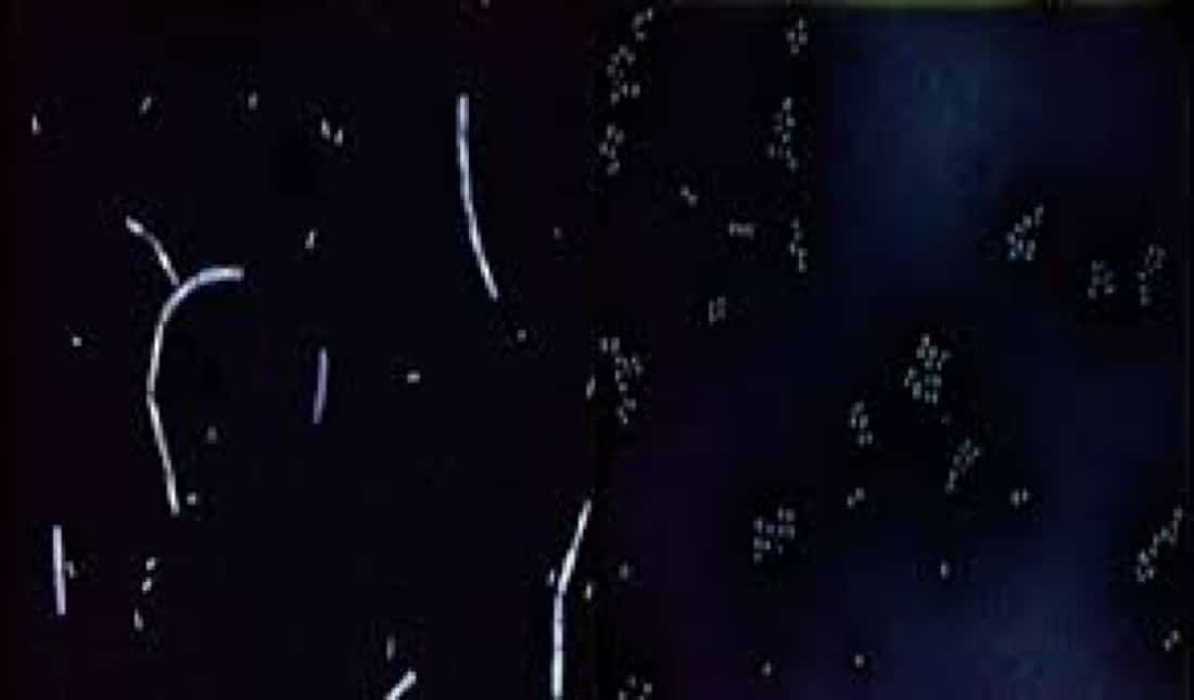
One of the most common negative staining agents is uranyl acetate, a uranium salt. Due to its ability to absorb electrons, uranyl acetate creates a dense, electron-dense background that surrounds the specimen. This contrast enhances the visibility of the specimen’s fine details, making it an ideal technique for observing delicate structures like viruses, bacteria, and cellular membranes.
Advantages of Negative Staining:
1. Preservation of Native Structure: Negative staining is particularly useful when preserving the native structure of a specimen is crucial. Traditional staining methods may alter the structure of the specimen, while negative staining allows for a more natural representation.
2. Visualization of Fine Structures: The high contrast provided by negative staining is especially beneficial for visualizing fine structures that might be challenging to observe with conventional staining techniques. This makes negative staining a valuable tool in fields such as virology, bacteriology, and structural biology.
3. Quick and Simple: Negative staining is a relatively quick and straightforward technique compared to many other staining methods. It requires minimal sample preparation, making it a practical choice for researchers looking to obtain rapid results.
Applications of Negative Staining:
1. Virology: Negative staining has been instrumental in the study of viruses, allowing scientists to observe the morphology and surface details of viral particles. This has contributed significantly to the understanding of viral structures and the development of antiviral treatments.
2. Bacteriology: The technique is widely used in bacteriology to study bacterial morphology and flagella. Negative staining aids in differentiating bacterial species and provides insights into their cellular structures.
3. Electron Microscopy: Negative staining is frequently employed in electron microscopy, where the technique enhances the contrast of biological specimens for detailed imaging at the subcellular level.
Challenges and Considerations:
While negative staining offers numerous advantages, it is not without its challenges. The technique may introduce artifacts, and the choice of staining agent and conditions must be carefully considered to avoid potential distortions in the specimen.
Conclusion:
Negative staining is a valuable technique in the realm of microscopy, offering researchers a powerful tool for visualizing delicate structures with enhanced contrast. Its applications span various scientific disciplines, contributing to our understanding of the microscopic world. As technology continues to advance, negative staining techniques are likely to evolve, providing even greater insights into the intricate details of biological specimens.


