Unraveling the Mysteries of Gel Electrophoresis: A Comprehensive Guide
Introduction:
In the realm of molecular biology and genetics, few techniques have had as profound an impact as gel electrophoresis. This powerful tool allows scientists to separate and analyze DNA, RNA, and proteins based on their size and charge. Since its inception in the 20th century, gel electrophoresis has become a cornerstone technique in laboratories worldwide, enabling breakthroughs in fields ranging from forensic science to medical diagnostics. This article aims to delve into the intricacies of gel electrophoresis, exploring its principles, methodologies, applications, and future prospects.
Principles of Gel Electrophoresis:
At its core, gel electrophoresis relies on the fundamental principles of electromagnetism and molecular biology. The process involves applying an electric field to a gel matrix, typically composed of agarose or polyacrylamide, submerged in a conductive buffer solution. The gel acts as a molecular sieve, impeding the movement of charged molecules based on their size and charge.
When a voltage is applied across the gel, negatively charged molecules, such as DNA fragments or proteins, migrate towards the positively charged electrode (anode) at a rate proportional to their size and charge. Smaller molecules move more quickly through the gel pores, while larger molecules experience greater resistance and migrate more slowly. As a result, molecules of different sizes become spatially separated within the gel matrix, forming distinct bands or patterns that can be visualized and analyzed.
Methodologies of Gel Electrophoresis:
Gel electrophoresis encompasses various methodologies tailored to specific applications and experimental requirements. The two primary types of gel electrophoresis are agarose gel electrophoresis and polyacrylamide gel electrophoresis (PAGE), each offering unique advantages and suitability for different molecular targets.
1. Agarose Gel Electrophoresis:
Agarose gel electrophoresis is widely used for the separation of DNA fragments, particularly in molecular biology research and genetic diagnostics. Agarose, derived from seaweed polysaccharides, forms a porous gel matrix when mixed with a buffer solution and allowed to solidify. The concentration of agarose in the gel determines its pore size and, consequently, the resolution of DNA fragments.
The process of agarose gel electrophoresis typically involves the following steps:
a. Preparation of the Gel: Agarose powder is mixed with a buffer solution, heated to dissolve the agarose, and then poured into a casting tray. Combs are inserted into the gel to create wells for loading samples.
b. Loading of Samples: DNA samples, often mixed with loading dye for visualization, are pipetted into the wells of the gel.
c. Electrophoresis: The gel is submerged in a buffer-filled chamber, and electrodes are connected to the gel tank. Upon applying a voltage, DNA molecules migrate through the gel according to their size, with smaller fragments traveling farther from the well.
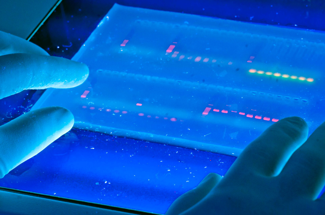
d. Visualization and Analysis: After electrophoresis, the gel is stained with a DNA-binding dye, such as ethidium bromide or SYBR Safe, and exposed to ultraviolet (UV) light for visualization of DNA bands. The size of DNA fragments is estimated by comparing their migration distance to molecular weight markers run alongside the samples.
2. Polyacrylamide Gel Electrophoresis (PAGE):
Polyacrylamide gel electrophoresis offers higher resolution and is commonly employed for the separation of proteins and small nucleic acids, such as RNA fragments and oligonucleotides. Polyacrylamide gels are formed through the polymerization of acrylamide monomers in the presence of a cross-linking agent, typically N,N’-methylenebisacrylamide.
The process of PAGE involves similar steps to agarose gel electrophoresis, with some key differences:
a. Gel Preparation: Polyacrylamide gels are cast with precise concentrations to achieve optimal resolution for the target molecules. Additionally, special reagents may be incorporated into the gel to facilitate protein denaturation or maintain RNA secondary structure.
b. Loading and Electrophoresis: Protein or nucleic acid samples are denatured, if necessary, and loaded onto the gel wells. During electrophoresis, proteins migrate based on their size and charge, while RNA molecules may also be separated according to their secondary structure.
c. Staining and Visualization: Protein bands on PAGE gels can be visualized using protein-specific stains, such as Coomassie Brilliant Blue or silver stain. For RNA, specialized stains like ethidium bromide or SYBR Gold may be used. Alternatively, proteins and nucleic acids can be transferred onto membranes for Western blotting or Northern/Southern blotting, respectively, enabling detection with specific antibodies or probes.
Applications of Gel Electrophoresis:
Gel electrophoresis finds widespread applications across various scientific disciplines, including molecular biology, genetics, forensics, clinical diagnostics, and biotechnology. Some of the key applications include:
1. DNA Fragment Analysis: Gel electrophoresis is routinely used to analyze DNA fragments generated through techniques such as PCR (polymerase chain reaction), restriction enzyme digestion, and DNA sequencing. It facilitates the characterization of DNA samples, including genotyping, DNA fingerprinting, and mutation detection.
2. Protein Separation and Analysis: PAGE enables the separation of proteins based on their size, charge, and molecular weight. This technique is fundamental to protein purification, characterization, and quantification, as well as the study of protein-protein interactions and post-translational modifications.
3. RNA Analysis: Gel electrophoresis plays a crucial role in the analysis of RNA molecules, including mRNA expression profiling, detection of splice variants, and assessment of RNA integrity. Additionally, specialized techniques like northern blotting and RNA mobility shift assays rely on gel electrophoresis for RNA visualization and analysis.
4. Clinical Diagnostics: Gel electrophoresis is utilized in clinical laboratories for the diagnosis and monitoring of genetic disorders, infectious diseases, and cancer. It enables the detection of specific DNA mutations, RNA biomarkers, and protein abnormalities associated with various diseases.
5. Forensic Science: Gel electrophoresis is instrumental in forensic DNA analysis, allowing the comparison of DNA profiles from crime scene evidence with those of suspects or databases. Techniques like short tandem repeat (STR) analysis and DNA profiling rely on the high resolution of gel electrophoresis for accurate identification.
Future Perspectives and Advancements:
Despite its longstanding prominence, gel electrophoresis continues to evolve with advancements in technology and methodology. Emerging trends and innovations in gel electrophoresis include:
1. Microfluidic Electrophoresis: Miniaturization and automation of electrophoresis systems using microfluidic devices offer enhanced speed, sensitivity, and throughput for nucleic acid and protein analysis. Microchip-based electrophoresis platforms enable rapid sample processing and integration with downstream applications, such as next-generation sequencing and single-cell analysis.
2. High-Throughput Analysis: Gel electrophoresis is increasingly being integrated into high-throughput screening and analysis workflows, enabling the simultaneous processing of multiple samples and the parallel analysis of diverse molecular targets. Automated gel imaging systems and software tools facilitate the rapid quantification and interpretation of gel electrophoresis data, streamlining research and diagnostics.
3. Multimodal Separation Techniques: Hybrid approaches combining gel electrophoresis with other separation techniques, such as cap
illary electrophoresis, chromatography, and mass spectrometry, are expanding the analytical capabilities and versatility of gel-based assays. These integrated platforms offer complementary advantages, including enhanced resolution, sensitivity, and specificity for complex sample analysis.
Conclusion:
Gel electrophoresis stands as a foundational technique in molecular biology and biotechnology, enabling the separation, characterization, and analysis of nucleic acids and proteins with remarkable precision. From basic research to clinical diagnostics and forensic investigations, gel electrophoresis continues to shape our understanding of biological processes and drive scientific discovery. As technology advances and new methodologies emerge, the future of gel electrophoresis promises even greater insights into the complexities of the molecular world.


