Unlocking Nature’s Defense: Understanding Endospore Staining
In the microscopic world, where battles for survival are waged unseen to the naked eye, bacteria employ ingenious strategies to endure harsh conditions. One such survival mechanism is the formation of endospores, tough and resilient structures that enable certain bacteria to withstand extreme temperatures, desiccation, and chemical assaults. Endospore staining, a specialized laboratory technique, unveils these hidden fortresses, shedding light on the resilience of these microorganisms.
What are Endospores?
Endospores are dormant, tough, and non-reproductive structures formed by certain bacterial species as a response to unfavorable environmental conditions. These conditions might include nutrient depletion, extremes of temperature, pH, or the presence of toxic chemicals. Rather than a reproductive mechanism, endospore formation serves as a survival strategy, allowing bacteria to withstand inhospitable environments until conditions improve.
Endospore Staining: Peering into Microbial Fortresses
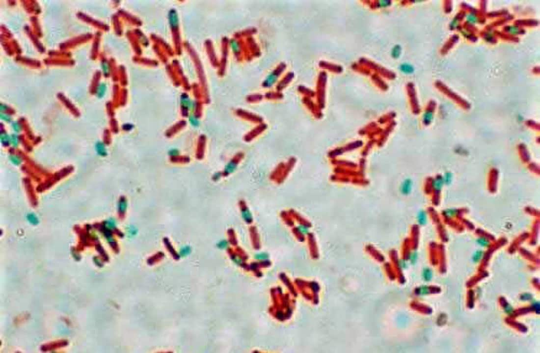
Endospore staining is a differential staining technique employed in microbiology laboratories to distinguish endospores from vegetative bacterial cells. Developed by the German bacteriologist Ferdinand Cohn in the 19th century, this technique utilizes specific dyes and heat to selectively colorize endospores, rendering them visible under the microscope.
The Schaeffer-Fulton Method: A Time-Tested Approach
The most widely used technique for endospore staining is the Schaeffer-Fulton method. This method involves the following steps:
1. Preparation of Smear: A bacterial smear is prepared on a clean glass slide from a pure culture of the organism under examination.
2. Heat Fixation: The smear is gently heat-fixed by passing it through a flame several times. Heat fixation not only attaches the bacterial cells to the slide but also kills them, preventing any potential contamination during staining.
3. Primary Stain: The smear is flooded with malachite green, the primary stain for endospores. Malachite green penetrates the endospore’s tough outer layer, staining it green.
4. Heat Treatment: The slide is gently heated to facilitate the penetration of the primary stain into the endospores. Heat also helps in softening the endospore coat, aiding dye uptake.
5. Decolorization: The slide is rinsed with water to remove excess stain. Then, the slide is flooded with water or a decolorizing agent such as water or alcohol. This step removes the primary stain from vegetative cells but not from endospores.
6. Counterstain: A contrasting counterstain, such as safranin or fuchsin, is applied to the smear. This stains the vegetative cells, imparting a contrasting color (usually pink or red) to them.
7. Washing and Drying: Excess counterstain is washed off, and the slide is allowed to air dry.
8. Examination: The prepared slide is examined under a light microscope. Endospores appear as green structures against a pink or red background, contrasting sharply with the stained vegetative cells.
Significance and Applications
Endospore staining is crucial for the identification and characterization of bacteria capable of forming endospores. It aids in the diagnosis of diseases caused by endospore-forming pathogens such as Bacillus anthracis (the causative agent of anthrax), Clostridium tetani (responsible for tetanus), and Clostridium botulinum (the bacterium behind botulism).
Moreover, endospore staining finds applications beyond medical microbiology. In industries such as food and beverage production, pharmaceuticals, and agriculture, where microbial contamination poses significant risks, the ability to detect endospores helps in implementing effective sanitation and sterilization protocols.
Conclusion
Endospore staining is a powerful technique that allows microbiologists to delve into the hidden world of microbial survival strategies. By selectively staining endospores, this method unravels the resilience of certain bacteria, offering insights into their ecological roles and pathogenic potential. As our understanding of microbial physiology and ecology continues to deepen, endospore staining remains a valuable tool in unraveling the mysteries of the microbial world.


