Staphylococcus Aureus 
Staphylococci are gram positive cocci that typically occur in grape-like clusters. Staphylococcus aureus is one of pathogen in humans normal formula.
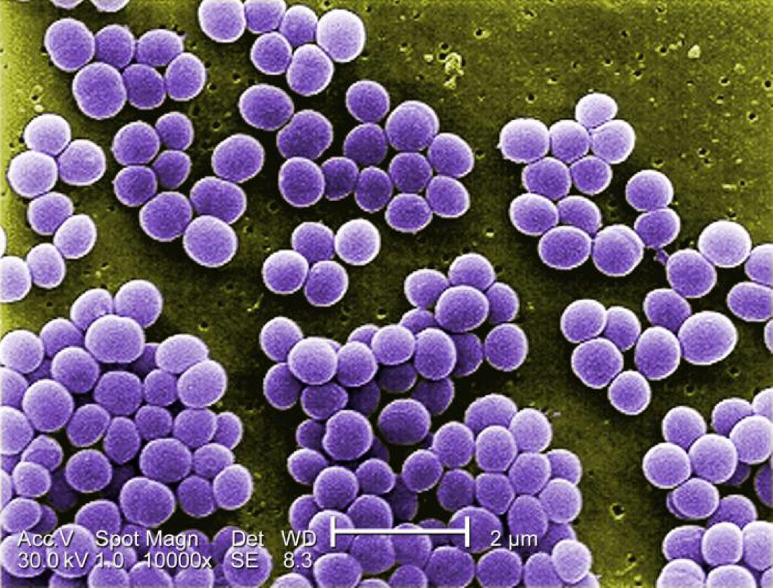
Morphology
They are spherical cocci, approximately 1 micrometer in diameter, arranged characteristically in grape-like clusters. These clusters are formed because of cell division occurring in three planes, with daughter cells tending to remain in close proximity.
In liquid culture they may also found single, in pairs and in short chains of three or four cells.
Staphylococcus aureus are non-motile and non sporing. A few strains possess microscopically visible capsules, particularly in young cultures.
Under the influence of penicillin and certain other chemicals, they may change to L forms.
Cultural Characteristics
Staphylococcus aureus grow readily on ordinary media within a temperature range of 10-42 C, the optimum being 37 C and pH 7.4 – 7.6. they are aerobes and facultative anaerobes.
Staph. aureus, when grown on nutrient agar, after incubation for 24hours, the colonies are large (2 – 4 mm diameter), circular, convex, smooth, shiny, opaque and easily emulsifiable. Most strains produce golden yellow pigment, though some may be white, orange or yellow. Pigment does not diffuse into the medium. Pigment production occurs optimally at 22 C and only in aerobic cultures.
Pigment production enhanced when 1% glycerol monoacetate or milk is incorporated in the medium.
On nutrient agar slope, the confluence growth presents a characteristic ‘oil-paint’ appearance. The colonies on blood agar are similar to those on nutrient agar.
Most strains are hemolytic, especially when incubated under 20 – 25 % carbon dioxide. Hemolysis is marked on rabbit or sheep blood and weak on horse blood agar.
They grow on MacConkey’s medium, producing smaller colonies that are pink due to lactose fermentation.
In liquid media, uniform turbidity is produced. Several selective media have been devised for isolating Staph. aureus from specimens such as feces containing other bacteria. These include media containing 8 – 10 % NaCl (salt-milk agar, salt broth), lithium chloride and tellurite (Ludlam’s medium), and polymyxin.
For primary isolation of Staph aureus, sheep blood agar is recommended. Human blood should not be used as it may contain antibodies or other inhibitors.
Biochemical Reactions
Staph aureus ferment a number of sugars, producing acid but no gas. Sugar fermentation is of no diagnosic value except for mannitol, which is usually fermented by staph aureus but not by other species.
They are catalase positive (unlike streptococci) and usually hydrolysis urea, reduce nitrites, liquefy gelatin and are MR and VP positive but indole negative. Most strains are lipolytic and when grown on media containing egg yolk, produce a dense opacity.
Production of phosphatase can be demonstrated by culturing on nutrient agar containing phenolphthalein diphosphate.
Staph aureus strains usually exhibit the following characteristics:
- Coagulase positive
- Greater biochemical activity, ferment mannite.
- Produce clear hemolysis on blood agar
- Produce a golden yellow pigment
- Liquefy gelatin
- Produce phosphatase
- Produce thermos table nucleases which can be demonstrated by the ability of boiled cultures to degrade DNA in an agar diffusion test.
Resistance
Staphylococci are among the more resistant of non sporing bacteria. if dried on threads, they remain their viability for 3 – 6 months. They have been isolated from dried pus after 2 – 3 months.
They may withstand 60 C for 30 minutes. Their thermal death point is 62 C for 30 minutes. Some staphylococci require 80 C for one hour to be killed.
Heat resistant strains have the ability to grow at a higher temperature, even at 45 C. most strains can grow in the presence of 10 % NaCl and some even in 15 % NaCl. These features are of significance in food preservation.
They resist 1% phenol for 15 minutes. Mercury perchloride 1% solution kills them in 10 minutes. Many aniline dyes are strongly bactericidal, crystal violet being lethal at a concentration of one in five hundred thousand and brilliant green, one in ten million.
Pathogenicity And Virulence
Staphylococci produce two types of diseases – infections and intoxications.
In the infection, the cocci gain access to damaged skin, mucosal or tissue sites, colonies by adhering to cells or extra cellular matrix, evade host defence mechanisms, multiply and cause tissue damage.
In intoxications, the disease is caused by the bacterial toxins produced either in the infected host or performed in vitro.
Staphylococcal Diseases
Because of presence in normal flora staphylococcal infections are among the most common of bacterial infections and range from the trivial to the fatal. Staphylococcal infections are characteristically localised pyogenic lesions, in contrast to the spreading nature of streptococcal infections. Common staphylococcal infections are as follow:
Skin and soft tissue: follicular is, furuncle (boil), abscess (particularly breast abscess), wound infection, carbuncle, impetigo, paronychia, less often cellulitis.
Musculoskeletal: Osteomyelitis, arthritis, bursitis, pyomyositis.
Respiratory: Tonsillitis, pharyngitis, sinusitis, otitis, bronchopneumonia, lung abscess, empyema, rarely pneumonia.
Central nervous system: abscess, meningitis, intracranial thrombophlebitis.
Endovascular: Bacteremia, septicemia, premika, endocarditis.
Urinary: Staphylococci are uncommon in routine urinary tract infections, though they cause infection in association with local instrumentation, implants or diabetes.
Staphylococcal disease may follow endogenous or exogenous infection. The mode of transmission may be by contact, direct or through fomites, by dust or by airborne droplets.
Hospital infections by staphylococci deserve special attention because of their frequency and because they are caused by strains which are resistance to many antibiotics.
Staphylococci are a common cause of postoperative wound infection and other hospital cross infections.
Laboratory Diagnosis
The specimens to be collected depend on the type of lesion (for example, pus from suppurative lesions, sputum from respiratory infections). In case of food poisoning, feces and the remains of suspected food should be collected.
Direct microscopy with Gram stained smears is useful in the case of pus, where cocci in clusters may be seen. This is of no value for the specimens like sputum where mixed bacterial flora are normally present.
Diagnosis may readily be made by culture. The specimens are plated on blood agar. Staphylococcal colonies appear after overnight incubation.
Specimens where staphylococci are expected to be scanty and outnumbered by other bacteria are inoculate on selective media like Ludlam’s or salt-milk agar or Robertson’s cooked meat medium containing 10% sodium chloride. Smears are examined from the cultures and the coagulase test done when staphylococci are isolated.
Antibiotic sensitivity tests should be performed as a guide to treatment. This is important as staphylococci readily develop resistance to drugs.
Bacteriophage typing may be done if the information is desired for epidemiological purposes.
Serological tests may sometimes be of help in the diagnosis of hidden deep infections.
Treatment
As drug resistance is so common among staphylococci, the appropriate antibiotic should be chosen based on antibiotic sensitivity test.
Benzyl penicillin is the most effective antibiotic, if the strain is sensitive.
Reference:
Text Book Of Microbiology


