Isolation Of Bacteriophage
Isolation Of Bacteriophage From Sewage
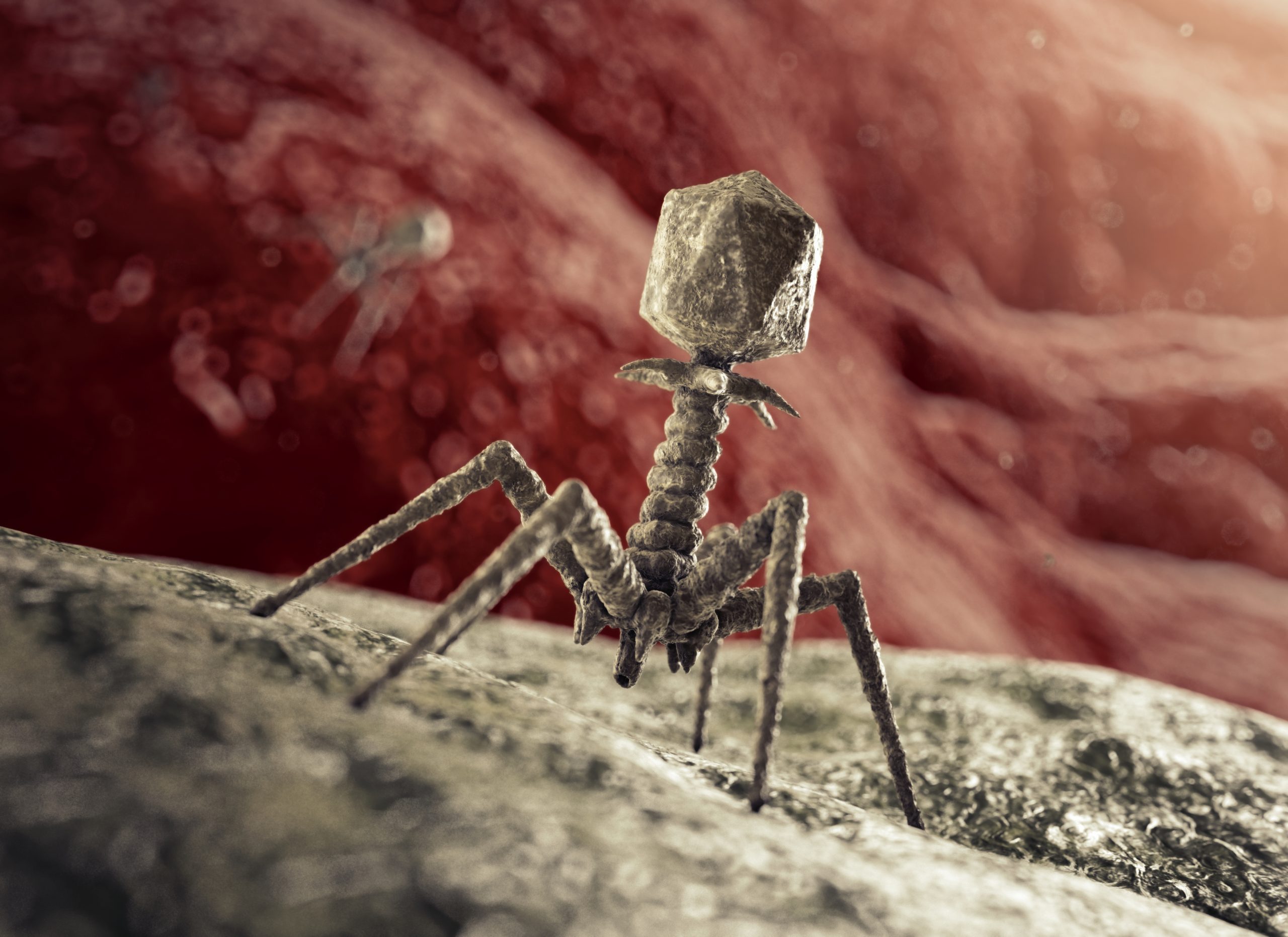
Although bacteriophages present in soil, sewage and even gut of mammals but isolation from these sources are difficult. The simple and easy way to siolate them is from raw sewage due to their presence in high concentrations. Most of the phages present in sewage infect E. coli and these are designated by a letter T, indicating the types. Seven types have been indentified and named as T-even phages (T2, T4, T6) and T-odd phages (T1, T3, T5 and T7). Both differs in size, form and other features but all are capable of infecting E. coli. There are three main steps for the isolation of bacteriophage from sewage. It is necessary to increase their number followed by separation from bacteria and finally to produce the plaques. The first process can be performed by enrichment with nutrients followed by filtration and finally seeding
with suitable bacteria. The sewage sample is the rich source of bacterial, viral and fungal pathogens. Hence, it is the first requirement for detection of bacteriophage.
Requirements
- Sewage
- Phage broth
- Pure culture of E. coli
- Incubator
- Centrifuge
- Membrane filtration unit
- Erlenmeyer flask
- Test tubes
- Nutrient broth
- Soft nutrient agar
- Hard agar
- Petri dishes
- Procedure
First step – Filtration process:
- Collect the raw sewage from sewage treatment plant through main hole.
- Mix the sewage (100 ml) with 10 ml deca strength phage broth (DSPB) and 10 ml of E.coli suspension.
- Incubate the mixture at 37ºC for 24 hours.
- Centrifuge the sewage at 2500 rpm for 10 minutes, so that the bacteria and other solid matrix are separated out. Decant the supernatant
- Repeat the process for three times to obtain the filtrate.
- Then filter it through membrane filter (0.45 mm) to remove the bacterial cells. It is quite possible that some times, filter gets clogged or choked, hence it is desirable to change the filter and pass the filtrate through new filter.
- Connect the filtration assembly through vacuum pump to quick the filtration process.
- Keep vacuum pump on so that filtrate may accumulate in flask of filtration unit.
- Transfer aseptically the final filtrate so obtained into a pre sterilized Erlenmeyer flask.
Second step – Seeding of E.coli in the filtrate:
- Procure 4 tubes of soft nutrient agar of jelly like consistency and keep on water bath at 50ºC. Labelling them as A, B, C, D.
- Pour 1 ml filtrate in tube A, 2 ml filtrate in tube B and 4 ml in tube C. Keep tube D blank having no filtrate.
- Inoculate all the tubes with 0.5 ml E. coli and pour into Petri dishes previously containing hard agar and label them A, B, C, and D respective to the tubes as shown in Fig. 12.1.
- After medium is solidified, invert the plates, keep them in incubator for several hours and examine for plaque formation.

Results
After incubation, if some plaques are formed record their diameter and see the changes occurring with incubation.
Reference
Dr. R. C. Dubey – Practical Microbiology


