Gram Staining: Revealing the Microbial World
Introduction
Gram staining is a fundamental microbiological technique that plays a pivotal role in identifying and classifying bacteria into two distinct groups: Gram-positive and Gram-negative. Developed by Danish bacteriologist Hans Christian Gram in 1884, this staining method has become a cornerstone in microbiology, aiding in the diagnosis and treatment of various infectious diseases and contributing to our understanding of bacterial biology. In this article, we will delve into the principles, procedure, significance, and applications of Gram staining.
Principles of Gram Staining
Gram staining relies on the differential cell wall structure of bacteria. Bacterial cell walls primarily consist of peptidoglycan, a complex mesh of sugars and amino acids. Gram-positive bacteria have a thick layer of peptidoglycan in their cell walls, while Gram-negative bacteria have a thinner peptidoglycan layer surrounded by an outer membrane containing lipopolysaccharides.
The Gram staining process involves four primary steps:
1. Fixation: Bacterial cells are first fixed onto a glass slide, usually through heat or chemical fixation. This step kills the bacteria and attaches them firmly to the slide.
2. Crystal Violet Staining: The fixed cells are then flooded with crystal violet, a purple-colored dye. Crystal violet binds to peptidoglycan in the bacterial cell walls, imparting a violet color to all cells.
3. Iodine Treatment: Next, iodine is applied, forming a crystal violet-iodine complex within the cells. This step stabilizes the crystal violet stain.
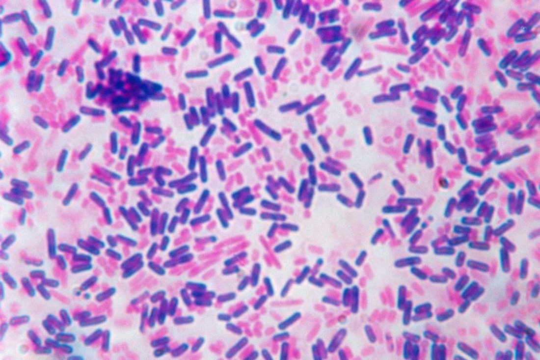
4. Decolorization: The critical step in Gram staining is the application of alcohol or acetone, which serves to remove the stain from some bacteria. Gram-positive bacteria retain the crystal violet-iodine complex due to their thick peptidoglycan layer, while Gram-negative bacteria lose the stain.
5. Counterstaining: To make the Gram-negative bacteria visible, a red counterstain, typically safranin, is applied. This stains the decolorized Gram-negative bacteria pink or red.
Procedure of Gram Staining
Gram staining is a relatively straightforward procedure when carried out with care. Here is a simplified step-by-step guide:
1. Prepare a bacterial smear on a clean glass slide and allow it to air dry.
2. Fix the smear by passing it briefly over a flame or using chemical fixatives.
3. Apply crystal violet for 1 minute, then rinse with water.
4. Add iodine solution and let it sit for 1 minute, followed by rinsing.
5. Carefully decolorize with alcohol or acetone until no more color washes off (usually 5-10 seconds).
6. Rinse immediately with water to stop the decolorization process.
7. Apply safranin for 1 minute, then rinse with water.
8. Blot the slide gently with a paper towel and allow it to air dry.
9. Examine the stained bacteria under a microscope, typically using oil immersion, if necessary.
Significance of Gram Staining
Gram staining is invaluable in microbiology for several reasons:
1. Bacterial Classification: It divides bacteria into Gram-positive and Gram-negative groups, aiding in the initial identification of unknown bacterial species.
2. Treatment Decisions: The Gram stain result can guide physicians in prescribing appropriate antibiotics. Gram-positive bacteria are generally susceptible to a wider range of antibiotics than Gram-negatives.
3. Infection Control: It helps identify potential pathogens in clinical settings, assisting in the prompt initiation of infection control measures.
4. Research and Taxonomy: Gram staining is a fundamental tool in bacterial taxonomy and research, providing insights into bacterial cell wall structure and evolution.
Applications of Gram Staining
The applications of Gram staining are widespread and include:
1. Clinical Microbiology: Identifying bacterial infections, determining the causative agent, and selecting the appropriate antibiotics.
2. Food Microbiology: Detecting spoilage or pathogenic bacteria in food products.
3. Environmental Microbiology: Studying microbial populations in soil, water, and air.
4. Pharmaceutical Industry: Quality control of drug products and assessing microbial contamination.
5. Research: Facilitating studies on bacterial morphology, antibiotic resistance, and bacterial physiology.
Conclusion
Gram staining is a cornerstone technique in microbiology that has stood the test of time. Its simplicity and effectiveness in distinguishing between Gram-positive and Gram-negative bacteria have made it an essential tool in various fields, from clinical medicine to scientific research. By revealing the microbial world in vivid hues of violet and pink, Gram staining continues to play a crucial role in our understanding and management of bacterial infections and the broader field of microbiology.


