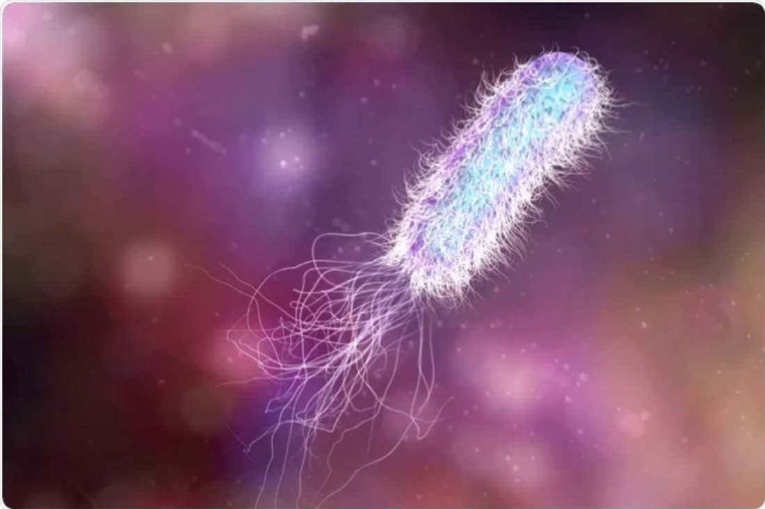
Flagella and Motility
Most motile procaryotes move by use of flagella (s. flagellum). Flagella is a threadlike locomotor appendages extending outward from the plasma membrane and cell wall. Here we would talk about bacterial flagella, those are best studied and explained.
Bacterial flagella are slender, rigid structures, about 20 nm across and up to 15 or 20 micrometer long. Flagella are so thin they cannot be observed directly with a bright-field microscope, but must be stained with special techniques designed to increase their thickness. The detailed structure of flagella can only be seen in electron microscope.
Bacterial species differ distinctively and their flagella structure too. Bacterial species can be identified by their flagella as well. Monotrichous bacteria (trichous means hair) have one flagellum, if it is located at end, it is said to be a polar flagellum. Amphitrichous bacteria (amphi means on both sides) have a single flagellum at each pole. In contrast locotrichous bacteria have a cluster of flagella at one or both ends. Peritrichous (peri means around) have flagella, spread fairly evenly over the whole surface of bacteria.
Flagella Ultrastructure
Transmission electron microscope studies have shown that the bacterial flagellum is composed of three parts. (1) the longest and most obvious portion is the flagella filament, which extends from the cell surface to the tip. (2) a basal body is embedded in the cell; and (3) a short, curved segment, the flagella hook, links the filament to its basal body and acts as a flexible coupling.
The filament is a hollow, rigid cylinder constructed of subunits of the protein flagellin, which range in molecular weight from 30,000 to 60,000 daltons, depending on the bacterial species.
The filament ends with a caping protein. Some bacteria have sheaths surrounding their flagella. For example, Bdellovibrio has a membranous structure surrounding the filament. Vibrio cholerae has a lipopolysaccharide sheath surrounding the filament.
The hook and basal body are quite different from the filament. Slightly wider than the filament, the hook is made of different protein subunits. The basal body is the most complex part of a flagellum. In E. coli and most gram-negative bacteria, the basal body has four rings connected to a central rod. The outer L and P rings associate with the lipopolysaccharide and peptidoglycan layers, respectively. The inner M ring contacts. The plasma membrane.
Gram – positive bacteria have only two basal body rings – an inner ring connected to the plasma membrane and an outer ring probably attached to the peptidoglycan.
The Mechanism Of Flagella Movement
Researchers have observed that prokaryotic flagella operate differently from eukaryotic flagella. The filament is in the shape of a rigid helix, and the cell moves when this helix rotates. Considerable evidence shows that flagella act just like propellers on a boat. Bacterial mutants with straight flagella or abnormally long hook regions cannot swim.

The nature of bacterial movement is determined by the direction of flagella rotation. Monotrichous, polar flagella rotate counter-clockwise (when viewed from outside the cell) during normal forward movement, whereas the cell itself rotates slowly clockwise. The rotating helical flagella filament thrusts the cell forward in a run with the flagellum trailing behind.
To move forward, the flagella rotate counterclockwise. As they do so, they bend at their hooks to form a rotating bundle that propels the cell forward. Clockwise rotation of the flagella disrupts the bundle and the cell tumbles.
Because bacteria swim through rotation of their rigid flagella, there must be some sort of motor at the base. A rod extends form the hook and ends in the M ring, which can rotate freely in the plasma membrane. It is thought that the S ring is attached to the cell wall in gram – positive cells and does not rotate. The P and L rings of gram – negative bacteria would act as bearings for the rotating rod. There is some evidence that the basal body is a passive structure and rotates within a membrane-embedded protein complex much like the rotor of an electrical motor turns in the center of a ring of electromagnets.
Reference: Microbiology by Prescott


