Dorner-staining method for bacterial endospores
Species at Bacillus Clostridium, Desulfotomaculum, Sporosarcina, etc. have the ability to form thick walled oval body. These cells have large amount of dipicolinic acid (5-10% of spore dry weight) which is undetectable in vegetative cells. Bacterial spores are best observed under phase contrast microscope. These appear as brilliant refractile body against phase dark cells.
Requirements
- Starved culture or spores
- Filter paper
- Carbol fuchsin
- Acid-alcohol
- Nigrosin Slide
- Cover slip
- Bunsen burner
- Inoculation needle
Procedure
(i) Prepare the smear on a slide and keep on it a paper saturated with carbol fuchsin stain (Fig. 3.5).
(ii) Pass the preparation through steam for 5-10 min.
(iii) Remove the paper and decolourise the film with acid-alcohol (3 ml conc. HCl in 97 ml, 95% ethanol) for 1 min, rinse with water and blot dry.
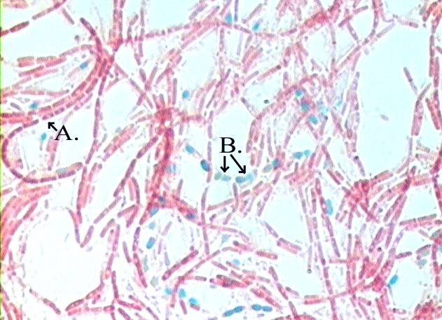
(iv) Place a drop of 7% nigrosin on the slide, cover it with another slide and draw the two apart from each other to form thin film of the nigrosin over the stained smear. The vegetative cell appears colourless while endospore appears red in colour under oil immersion objective.

Reference
Dr. R. C. Dubey – Practical Microbiology


