Yersinia Pestis: Morphology, Cultural Characteristics, Biochemical Reactions, Resistance, Diagnosis
The plague bacillus and many other Gram negative, short bacilli that are primary pathogens of rodents were grouped together in the genus Pasteurella. On the basis of cultural and biochemical differences, this group has been devided into three genera – Yersinis, Pasteurella and Francisells.
The genus Yersinia, containing the medically important species Y. pestis, the causative agent of plague.
The plague bacillus was discovered independently and simultaneously by Yersin and Kitasato (1894) in Hong Kong.
Morphology
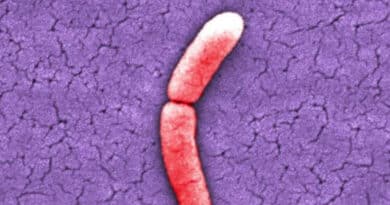
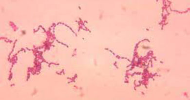
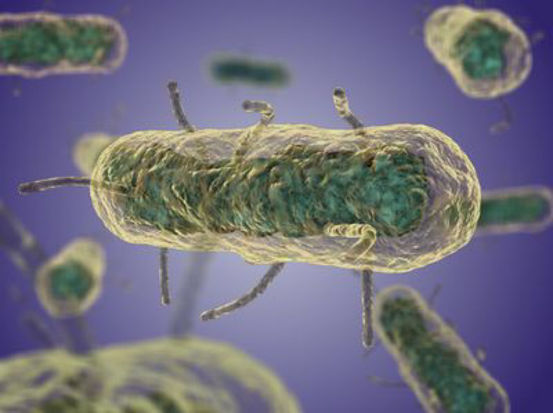
Y. pestis is a short, plump, ovoid, Gram negative bacillus, about 1.5 x 0.7 micrometer size, with rounded ends and convex sides, arranged singly, in short chains or in small groups.
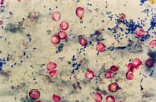
In smears stained with Giemsa or methylene blue, it shows bipolar staining (safety pin appearance) with the two ends densely stained and the central area clear.
The bacillus is surrounded by a slime layer (envelope or capsule). It is nonmotile, non sporing and non acid fast.
Cultural Characteristics
Y. pestis is aerobic and facultatively anaerobic. Growth occurs over a wide range of pH (pH 5- 9.6, optimum pH 7.2) and temperature (range 2 – 45 C). the optimum temperature for growth (unlike most pathogens) is 27 C but the envelope develops best at 37 C.
It grow well on ordinary media. On nutrient agar, colonies are small, delicate, transparent discs, becoming opaque on continued incubation.
Its colonies of blood agar or other hemin containing media are dark brown due to the absorption of the hemin pigment. Colorless colonies are formed on MacConkey’s agar. In broth, a flocculent growth occurs at the bottom and along the sided of the tube, with little or no turbidity.
Biochemical Reactions
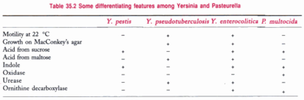
Glucose, maltose, and mannitol but not lactose, sucrose or rhamnose are fermented with the production of acid but no gas. Indole is not produced. It is MR positive and VP and citrate negative, catalase positive and aesculin positive and oxidase and urease negative. Gelatin is not liquefied.
Resistance
The plague bacillus is easily destroyed by exposure to heat, sunlight, drying and chemical disinfectants. It is destroyed by heat and chemical disinfectants. It is destroyed by heat at 55 C or by 0.5% phenol in 15 minutes. It remains viable for long periods in cold, moist environments. It can survive for several months, and even multiply, in the soil of rodent burrows. All strains are lyses by a specific antiplague bacteriophage at 22 C.
Laboratory Diagnosis
The laboratory should be able to diagnose plague not only in humans, but in rodents also, as timely detection of infection in rats may help to prevent epidemic spread.
A rat which died of plague may carry infected fleas and should be handled with care. Pouring kerosene oil over the carcass is a simple method of eliminating the fleas. In the laboratory, the carcass should be dipped in 3% lysol to destroy ectoparasites.
During epizootics, it is easy to diagnose plague in rats. Buboes are usually present in the cervical region. They are hard and can be moved under the skin. On section, the bubo may show congestion, hemorrhagic points or grey necrosis. Smears from the bubo stained with methylene blue show the bipolar bacilli.
The fluorescent antibody technique may be of use in identifying plague bacilli in the impression films of the tissues.
Reference: The Text Book Of Microbiology