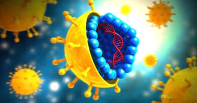
Cultivation Of Plant Virus
Plant virus can be cultivated by direct mechanical Inoculation of virus suspension by rubbing on leaves of living plants. This can lead to formation of local lesions as well as general infection. Some plant virus can replicate to large numbers in infected plants. For example, a single hair cell of an infected tobacco plant may contain over 10^7 tobacco mosaic visions. Indeed, as much as 10 percent of the dry weight of infected leaves may be tobacco mosaic virus. Transfer of infection from cell to cell occurs mainly by direct transfer of various via plasmodesmata.
Effects of virus infection on cells
Disease symptoms in the host due to virus infection vary from None to massive destruction of infected cells leading to cell death. In cell tissue culture, groups of killed cells (plaques) have been used in the enumeration of viruses because the number of plaques is proportional to the number of infectious virus particles present. Each virus gives rise to a single plaque, just as a bacterium gives rise to a single colony.
Other effects of cell infection by viruses include formation of giant cells (polykaryotes), creation of genetic changes such as chromosomal breakage, induction of interferon production by the infected cell that prevents infection of healthy cells, appearance of pocks or necrotic lesions on the chorioallantoic membrane of embryonated eggs, and formation of inclusion bodies.
Inclusion bodies
Before it was possible to study the morphology of viruses at the high magnifications provided by the electron microscope, investigators using light microscopy had observed intracellular structures, or inclusion bodies, associated with virus diseases.
J. B. Buist noted small particles in the cytoplasm of cells surrounding the lesions of smallpox. These he called elementary bodies. E. Patches made the same observation independently in 1906. It is known that these patches bodies are aggregates, or colonies of various growing in the cytoplasm of the host cell. In 1892, G. Garnier exported having seen small round particles in the cytoplasm of similar cells. These Guarnieri bodies are also thought to consist of aggregates of unassembled virus subunits and infect virjons.
Characteristic inclusion bodies are found in the cytoplasm of the purkinjie cells of the cerebellum and in certain other nerve cells in cases of rabies infection. Finding these typical inclusions (called Negri bodies after discoverer) is diagnostic of the disease.
Inclusion bodies are for the most part characteristic of the virus causing the infection and even suggest definite pathological changes in the cell. It is generally true, however, that inclusion bodies are aggregates of unassembled virus subunits and intact visions in infected cells. It is experimentally possible to remove inclusion bodies from the cell Ans use them as inoculum to infect other cells.