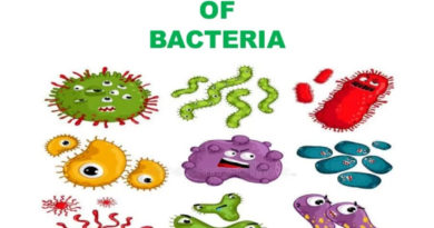
Gram’s Staining for differentiation of bacteria
Gram’s staining is one of the most important differential techniques used in bacteriology. There are two groups: gram positive and gram negative bacteria.
Method of gram staining was developed by “Christian Gram” in 1984.
In this method smear is stained with crystal violet and then iodine is applied as a moderate. The crystal violet iodine complex impart purple black color to the cells. In gram-positive cells this complex binds to the magnesium-ribonucleic acid component of the cell wall, forming a complex which is difficult to remove. The intensely stained cells are then washed with ethanol. This serves as a lipid solvent and a dehydrating agent for protein.
The gram positive bacteria contain low lipid content in their cell wall hence low amount of lipid is dissolved by alcohol. This makes minute pores in the cell wall that are closed by the dehydration effect of alcohol.
In gram negative cells, large pores are formed that do not close appropriately hence, dehydration of cell wall protein does not occur completely. So an unbound crystal violet complex leaves the cell colorless or unstained.
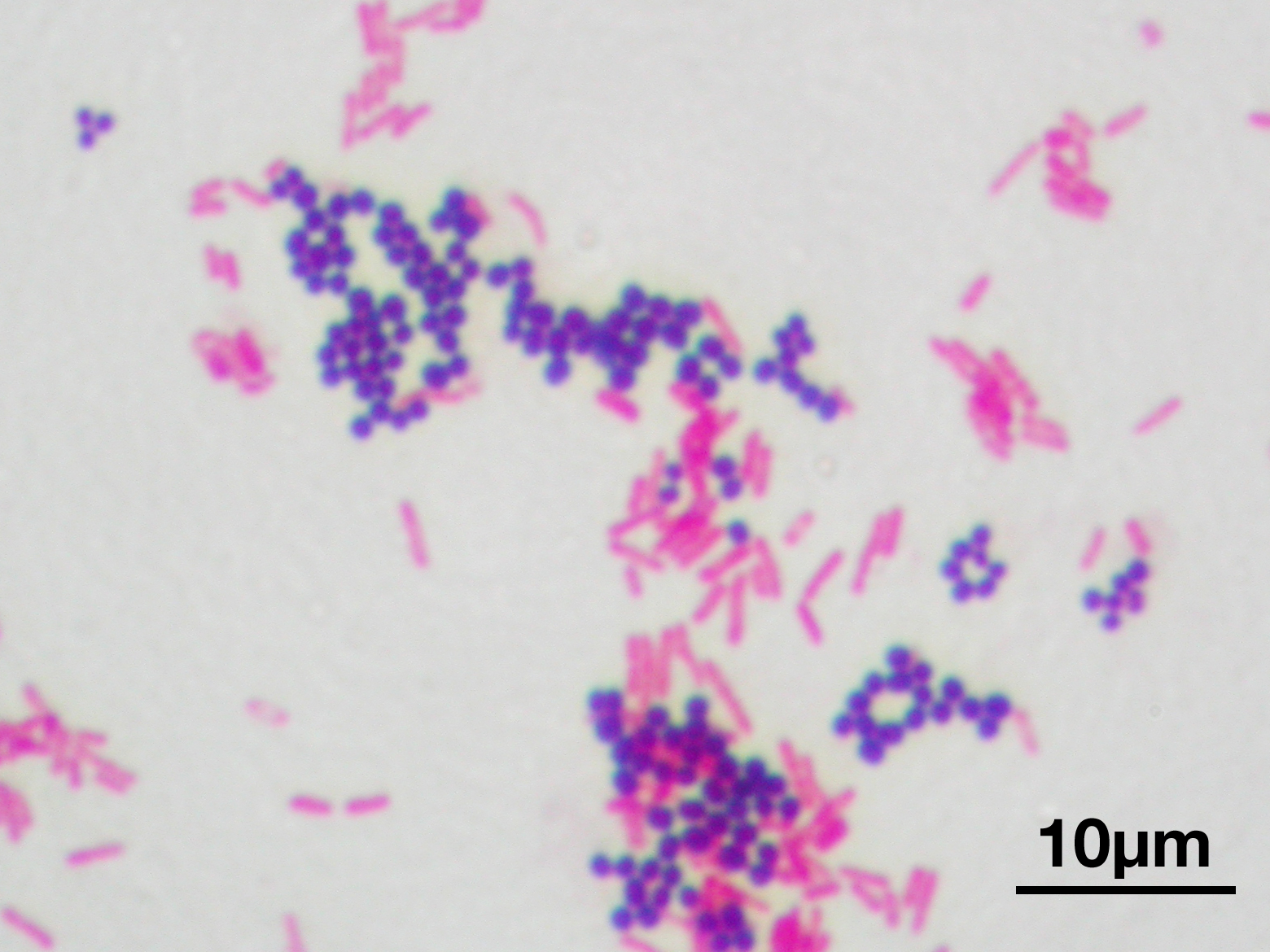
Now if the smear is counter stained with safranin, the gram negative cells are easily seen due to absorption of safranin and imparting the cells red color. While gram positive cells retain the blue color of the primary stain.
Requirements
- Bacterial slant culture
- Crystal violet
- Gram’s iodine
- Ethyl alcohol
- Safranin
- Bunsen burner
- Inoculation needle
- Staining tray
- Glass slide
- Microscope
Procedure
It is advisable to use young culture for gram staining.
- Prepare a bacterial smear and heat fixed on the slide using standard procedure. Pour a few drops of crystal violet on the smear.
- Wait for 1 minute and wash with tap water. Now, flood the smear with gram’s iodine for 1 minute and again wash with tap water.
- Decolourise the Staub with ethyl alcohol (95%) by dropping the reagent drop wise until crystal violet fails to wash from the smear.
- Wash it with tap water and counterstain with safranin for 45 seconds and wash again with water.
- After drying, examine under oil immersion.
Results
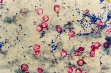
The bacteria that take stains and appear dark violet or blue black colored are called gram positive bacteria. Those appearing pink or red colored and do not stain are called gram negative bacteria.
Reference:
Dr. R C Dubey – Practical Microbiology